All living mammals (except humans) that have become true bipeds have adopted a very specific and specialized bounding form of bipedalism. Bipedal hopping results in a distinctive pelvic structure and this structure has similarities to specialized fossorial groups, implying that these disparate groups are incurring similar locomotor stressors in their respective modes of life.
Hoppers :
With regards to the pelvis, kangaroos seem to have adapted to a bipedal hopping way of life by changing elements of the pelvis such as the large pubis and ischium and the long narrow ilium.
They seem to share this pelvic morphology with other groups of unrelated mammal which also employ a bounding type of locomotion and also with groups that are fossorial.
Pelvis of kangaroos and kin:
A boxy pelvis with well developed pubis, ischiopubic ramus and ischium. Also a long, narrow, almost pronglike ilium.


tree kangaroo

Some hopping rodents:

Springhare

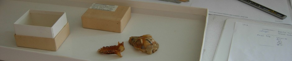
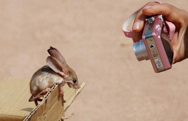
jerboa
Diggers:
This morphology of the pelvis appears again in mammal groups which are highly fossorial. The elongated, winglike ilium, and an overall boxy pelvis formed by a robust pubis and ischium, implys that some of the same stresses inherent in flexing and extending the hip during the bound are also acting in an animal that extends the limb backword against a substrate (i.e. digging)
cape dune mole rat

Ground squirrel

Cane Rat

Muscles of the Pelvis:
Published knowledge of muscular and osteology of the pelvis and hind limb of macropods seem to be lacking, with the exception of Hopwood 1976.
According to Hopwood, the biceps femoris, obturator exertenis, gemmelli, and iliopsoas are all fleshy well-developed muscles with origins on the pelvis.
The biceps femoris, part of the hamstrings group, aids in knee flexion and hip extension and stabilizes the hip. The obturator externis and gemellus both help stabilize the hip joint during locomotion, and the iliopsoas group aids in flexion of the thigh.
(PAUL R. HOPWOOD AND REX M. BUTTERFIELD. The musculature of the proximal pelvic limb of the Eastern Grey Kangaroo Macropus major (Shaw) Macropus giganteus (Zimm). J. Anat. (1976), 121, 2, pp. 259-277 )

LENGTHS OF PELVIC ELEMENTS:
Ischium length has been correlated with locomotor mode in old world monkeys, being shorter in the leaping monkeys like the colobus (more acceleration), longer in the more terrestrial cercopithecines. A shorter ischium gives greater acceleration in the leap, while a longer ischium gives greater hamstring power.
(K. Steudel. 1981. Functional Aspects of Primate Pelvic Structure: A Multivariate Approach. AMERICAN JOURNAL OF PHYSICAL ANTHROPOLOGY 55:399-410 )
While data on ishium and pubic length is lacking to non-existent outside of primates, the well-developed ischium and pubis in macropods, leaping rodents, and fossorial rodents, suggests a need for strong hamstring muscles and muscle power rather than fast acceleration.
Which absolutely begs the question:
Who would win a standing start, a kangaroo or a Bugatti Veyron Super Sport?


http://ru.wikipedia.org/wiki/%D0%A4%D0%B0%D0%B9%D0%BB:Skeleton_of_Cape_Dune_Mole_Rat.jpg
http://snakephotographer.com/?tag=jerboa
http://www.thefeaturedcreature.com/2010_11_28_archive.html#axzz24Kfgnq9j
(http://www.tree-kangaroo.net/tkInfo.html)
http://www.nhc.ed.ac.uk/index.php?page=24.134.165.255.266
http://www.flickr.com/photos/26485684@N08/2885093922/
http://www.zoology.wisc.edu/uwzm/collections.html
http://www.spcollege.edu/hec/vt/vtde/anatomy/3a.htm
http://vanat.cvm.umn.edu/run/plate3.html
http://www.rightpet.com/Small-exotic-mammalDetail/greater-cane-rat
(Hudson, Penny E.; Corr, Sandra A.; Payne-Davis, Rachel C.; Clancy, Sinead N.; Lane, Emily; Wilson, Alan M.Functional anatomy of the cheetah (Acinonyx jubatus) hindlimb Journal of Anatomy, Apr2011, Vol. 218 Issue 4, p363-374)
UW Radiology. http://www.rad.washington.edu/academics/academic-sections/msk/muscle-atlas/lower-body/obturator-externus
http://www.animalspot.net/gallery/kangaroo-pictures
http://www.museumofosteology.org/museum-exhibits/5/Adaptation-Locomotion.htm


 According to Merck at the UM Dept. Geology the basal amniotes did have this kind of rudimentary tooth differentiation, kind of proto-canines. You can see it in Paleothyris, the earliest anapsid, Petrolacosaurus, the earliest diapsid, and Ophiacodon, an early synapsid. This trait seems to be imbedded deep in the roots of these two groups.


